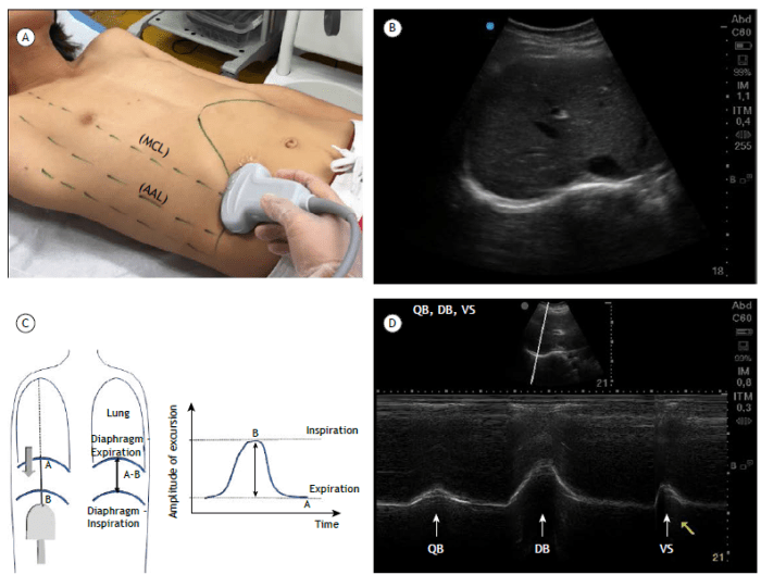
Crus of the diaphragm ultrasound – Ultrasound examination of the crus of the diaphragm provides invaluable insights into the anatomy, function, and pathology of this critical structure. This comprehensive guide delves into the sonographic appearance, clinical applications, and differential diagnosis of crus abnormalities, empowering clinicians with the knowledge to accurately assess and manage diaphragmatic disorders.
The crus of the diaphragm, composed of muscular fibers innervated by the phrenic nerve, plays a crucial role in respiration and abdominal compartmentalization. Ultrasound imaging enables visualization of the crus, facilitating the detection of hernias, tears, and other pathological conditions that may impair its function.
Define the crus of the diaphragm: Crus Of The Diaphragm Ultrasound
The crus of the diaphragm is a muscular structure that forms the lateral and posterior boundaries of the diaphragm, a dome-shaped muscle separating the thoracic and abdominal cavities. It plays a crucial role in respiration by contracting and relaxing to facilitate breathing.
Anatomical Location and Structure
The crus of the diaphragm is located on either side of the vertebral column, extending from the lumbar vertebrae (L1-L4) to the xiphoid process of the sternum. It consists of two distinct portions: the right crus and the left crus.
- Right crus:Arises from the bodies and intervertebral discs of the lumbar vertebrae (L1-L3) and inserts onto the right side of the central tendon of the diaphragm.
- Left crus:Arises from the bodies and intervertebral discs of the lumbar vertebrae (L1-L4) and inserts onto the left side of the central tendon of the diaphragm. The left crus is slightly longer and more robust than the right crus.
Muscular Composition and Innervation
The crus of the diaphragm is composed of skeletal muscle fibers innervated by the phrenic nerve. The phrenic nerve originates from the cervical spinal cord (C3-C5) and descends through the chest cavity to reach the diaphragm.
- Right crus:Innervated by the right phrenic nerve.
- Left crus:Innervated by the left phrenic nerve.
Describe the sonographic appearance of the crus of the diaphragm
The sonographic appearance of the crus of the diaphragm can vary depending on the imaging plane and the transducer frequency used. However, some general characteristics can be described.
Echogenicity
The crus of the diaphragm typically appears as a thin, hyperechoic (brighter than surrounding tissues) structure on ultrasound. This is due to the dense fibrous tissue that makes up the crus.
Thickness
The thickness of the crus of the diaphragm can vary, but it is typically around 2-3 mm. The thickness may be increased in certain conditions, such as obesity or cirrhosis.
Margins
The margins of the crus of the diaphragm are typically well-defined and smooth. However, they may be irregular in certain conditions, such as diaphragmatic hernias or tears.
Variations and Abnormalities, Crus of the diaphragm ultrasound
There are several variations and abnormalities that may be encountered when evaluating the crus of the diaphragm on ultrasound. These include:
- Diaphragmatic hernias: These are openings in the diaphragm that allow abdominal contents to herniate into the chest cavity. Diaphragmatic hernias can be congenital (present at birth) or acquired (develop later in life). They can be asymptomatic or cause a variety of symptoms, depending on the size and location of the hernia.
- Diaphragmatic tears: These are tears in the diaphragm that can be caused by trauma or surgery. Diaphragmatic tears can be partial or complete, and they can be asymptomatic or cause a variety of symptoms, depending on the size and location of the tear.
- Diaphragmatic eventration: This is a condition in which the diaphragm is thin and weak, causing it to bulge into the chest cavity. Diaphragmatic eventration is usually asymptomatic, but it can cause shortness of breath or other respiratory problems in severe cases.
Clinical applications of crus of the diaphragm ultrasound
Ultrasound evaluation of the crus of the diaphragm plays a significant role in various clinical scenarios, aiding in the diagnosis, monitoring, and guidance of interventions related to the diaphragm.
Diaphragmatic Paralysis
Ultrasound is a valuable tool for diagnosing and monitoring diaphragmatic paralysis. It allows visualization of the diaphragm’s movement and thickness, which can be impaired in paralysis.
Diaphragmatic Eventration
Ultrasound can detect diaphragmatic eventration, a condition characterized by an elevated diaphragm. It helps assess the extent of eventration and its impact on lung function.
Diaphragmatic Herniation
Ultrasound is useful in diagnosing and monitoring diaphragmatic herniation, where abdominal contents protrude through an opening in the diaphragm. It allows visualization of the herniated structures and evaluation of their size and location.
Guidance for Interventions
Ultrasound can guide interventions such as phrenic nerve stimulation or surgical procedures involving the crus of the diaphragm. It provides real-time visualization of the target structures, ensuring accurate placement of electrodes or surgical instruments.
Differential diagnosis of crus of the diaphragm abnormalities

The differential diagnosis of crus of the diaphragm abnormalities includes a wide range of conditions that can cause abnormal ultrasound findings. These conditions can be broadly classified into hernias, tears, tumors, and inflammation.
Hernias
- Hiatal hernia:This is the most common type of hernia that can affect the crus of the diaphragm. It occurs when the stomach protrudes through an opening in the diaphragm called the esophageal hiatus. Hiatal hernias can be asymptomatic or can cause symptoms such as heartburn, regurgitation, and chest pain.
- Morgagni hernia:This is a rare type of hernia that occurs through the foramen of Morgagni, which is located between the costal and sternal attachments of the diaphragm. Morgagni hernias can contain abdominal contents such as omentum, small intestine, or colon.
- Bochdalek hernia:This is a congenital hernia that occurs through a defect in the posterior diaphragm. Bochdalek hernias can be life-threatening in newborns and can cause respiratory distress and other complications.
Tears
- Traumatic tears:These tears can occur as a result of blunt or penetrating trauma to the chest or abdomen. Traumatic tears of the crus of the diaphragm can be associated with other injuries, such as rib fractures or lung contusions.
- Iatrogenic tears:These tears can occur as a result of medical procedures, such as laparoscopic surgery or thoracentesis. Iatrogenic tears of the crus of the diaphragm are typically small and asymptomatic, but they can sometimes lead to herniation.
Tumors
- Benign tumors:These tumors are rare and can include lipomas, schwannomas, and neurofibromas. Benign tumors of the crus of the diaphragm are typically asymptomatic and do not require treatment.
- Malignant tumors:These tumors are also rare and can include sarcomas, lymphomas, and mesotheliomas. Malignant tumors of the crus of the diaphragm can be aggressive and may require surgery, chemotherapy, or radiation therapy.
Inflammation
- Diaphragmatitis:This is a condition characterized by inflammation of the diaphragm. Diaphragmatitis can be caused by a variety of factors, including infection, trauma, and autoimmune disorders. Diaphragmatitis can cause pain, shortness of breath, and other symptoms.
Imaging protocols for crus of the diaphragm ultrasound

To ensure a comprehensive evaluation of the crus of the diaphragm, an optimized ultrasound imaging protocol is essential. This protocol encompasses transducer selection, scanning planes, patient positioning, and image optimization techniques.
Transducer selection
- High-frequency linear array transducer (e.g., 5-12 MHz) provides excellent resolution for detailed visualization of the crus.
- Curvilinear array transducer (e.g., 2-5 MHz) offers a wider field of view, enabling assessment of the crus in a larger area.
Scanning planes
- Sagittal plane:Assesses the thickness, echogenicity, and continuity of the crus.
- Axial plane:Evaluates the cross-sectional area and shape of the crus.
Patient positioning
- Supine position with knees slightly flexed.
- Head turned to the contralateral side for better acoustic access to the diaphragm.
Image optimization
- Adjust gain and frequency settings to optimize image quality.
- Use harmonic imaging to enhance tissue contrast and reduce noise.
- Employ color Doppler to assess blood flow within the crus.
Reporting and documentation of crus of the diaphragm ultrasound findings

Reporting crus of the diaphragm ultrasound findings requires a structured approach to ensure clarity, accuracy, and completeness. A standardized reporting template can facilitate efficient and effective documentation of the examination findings.
Structured reporting template
- Patient demographics:Name, age, sex, relevant medical history
- Indication for examination:Suspected diaphragmatic injury, hernia, or other pathology
- Ultrasound equipment:Transducer type, frequency, settings
- Crus of the diaphragm measurements:Thickness (in mm) and length (in cm) of the right and left crus
- Echogenicity:Hyperechoic, hypoechoic, or isoechoic compared to adjacent muscle
- Margins:Well-defined, ill-defined, or disrupted
- Abnormalities detected:Hernias, tears, thickening, thinning, calcifications
Examples of clear and concise reporting language
“The right crus of the diaphragm measures 5 mm in thickness and 10 cm in length. It is hyperechoic compared to the adjacent liver parenchyma. The margins are well-defined, and no abnormalities were detected.”
“The left crus of the diaphragm measures 3 mm in thickness and 8 cm in length. It is hypoechoic compared to the adjacent spleen. The margins are ill-defined, and a small hernia is present at the posterolateral aspect.”
FAQ Resource
What are the characteristic ultrasound findings of a normal crus of the diaphragm?
A normal crus of the diaphragm appears as a thin, hyperechoic structure with well-defined margins.
What are the common causes of abnormal ultrasound findings in the crus of the diaphragm?
Common causes include hernias, tears, tumors, and inflammation.
What is the role of ultrasound in evaluating diaphragmatic paralysis?
Ultrasound can detect the absence of diaphragmatic motion, indicating paralysis.
How does ultrasound guide interventions involving the crus of the diaphragm?
Ultrasound can guide needle placement for diagnostic procedures and surgical repairs.