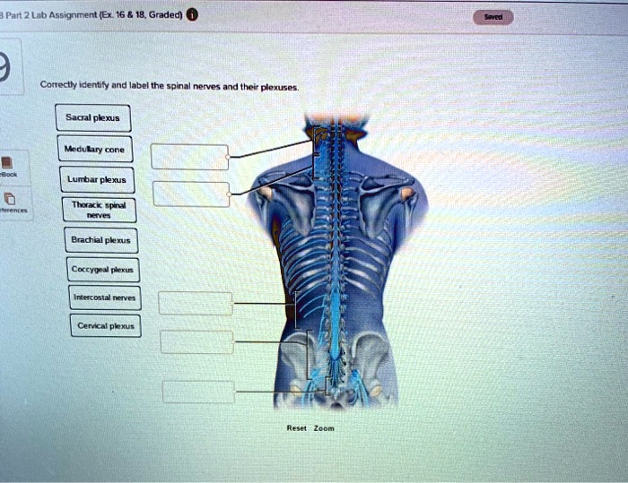
Correctly identify and label the spinal nerves and their plexuses. – Correctly identifying and labeling spinal nerves and their plexuses is crucial for understanding the intricate anatomy and function of the nervous system. This comprehensive guide delves into the significance of accurate identification, the formation and structure of spinal nerves, the composition of spinal plexuses, their clinical implications, and advanced techniques used for visualization and assessment.
Spinal nerves, originating from the spinal cord, play a vital role in transmitting sensory and motor information throughout the body. Spinal plexuses, formed by the merging of spinal nerves, innervate specific regions of the body, enabling movement, sensation, and autonomic functions.
Misidentification or mislabeling of these structures can have severe consequences for surgical procedures, pain management, and neurological assessments.
Introduction

Correctly identifying and labeling spinal nerves and their plexuses is of paramount importance in understanding the intricate anatomy and function of the nervous system. The spinal cord, the primary conduit of communication between the brain and the rest of the body, gives rise to 31 pairs of spinal nerves that branch out to innervate various parts of the body.
Accurate labeling of these nerves and their associated plexuses is essential for surgical procedures, pain management, and neurological assessments.
Spinal Nerves: Correctly Identify And Label The Spinal Nerves And Their Plexuses.

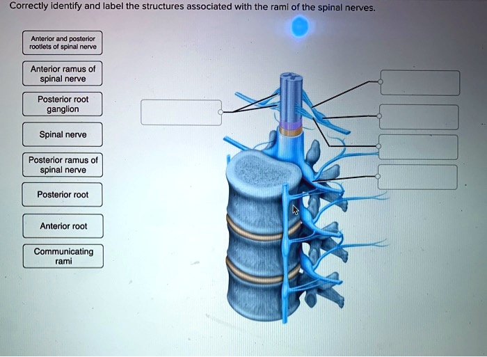
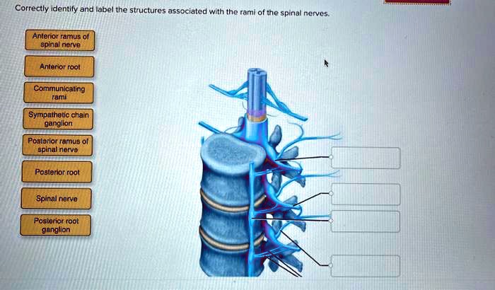
Spinal nerves are mixed nerves, meaning they contain both sensory and motor fibers. They are formed by the union of a dorsal root, which carries sensory information from the body to the spinal cord, and a ventral root, which carries motor commands from the spinal cord to the muscles.
The 31 pairs of spinal nerves are divided into five regions based on their location along the spinal cord:
- 8 cervical nerves (C1-C8)
- 12 thoracic nerves (T1-T12)
- 5 lumbar nerves (L1-L5)
- 5 sacral nerves (S1-S5)
- 1 coccygeal nerve (Co1)
Each spinal nerve innervates a specific area of the body, known as its dermatome and myotome. The dermatome refers to the area of skin supplied by the sensory fibers of the nerve, while the myotome refers to the muscles innervated by its motor fibers.
Spinal Plexuses

Spinal plexuses are networks of interconnected spinal nerves that form in the neck, chest, abdomen, and pelvis. These plexuses give rise to the peripheral nerves that innervate the limbs and other structures.
There are four major spinal plexuses:
- Cervical plexus (C1-C5): Innervates the neck, shoulders, and upper limbs
- Brachial plexus (C5-T1): Innervates the arms and hands
- Lumbar plexus (L1-L4): Innervates the lower abdomen, buttocks, and legs
- Sacral plexus (L4-S4): Innervates the pelvic organs, buttocks, and legs
Clinical Significance

Misidentifying or mislabeling spinal nerves and plexuses can have serious clinical consequences. During surgical procedures, accurate identification of nerves is crucial to avoid nerve damage and ensure proper function. In pain management, understanding the innervation patterns of spinal nerves helps guide nerve blocks and other pain-relieving interventions.
Case studies have demonstrated the importance of accurate nerve identification. For example, misidentification of the median nerve during carpal tunnel surgery can lead to persistent numbness and weakness in the hand. Similarly, mislabeling the sciatic nerve during hip replacement surgery can result in foot drop and other neurological deficits.
Advanced Techniques
Advanced imaging techniques play a vital role in visualizing and assessing spinal nerves and plexuses. Magnetic resonance imaging (MRI) and computed tomography (CT) scans provide detailed cross-sectional images of the spine and surrounding structures, allowing for precise identification of nerves and plexuses.
Electromyography (EMG) is another valuable technique that measures the electrical activity of muscles. By stimulating specific nerves and recording the muscle response, EMG can help diagnose nerve damage or dysfunction.
- MRI: Provides high-resolution images of the spine and surrounding tissues, including nerves and plexuses.
- CT scans: Offer detailed cross-sectional images of the spine and can help visualize bony structures that may compress nerves.
- EMG: Measures the electrical activity of muscles, helping diagnose nerve damage or dysfunction.
FAQ
What are the 31 pairs of spinal nerves?
The 31 pairs of spinal nerves are classified according to their level and region of the spinal cord: 8 cervical, 12 thoracic, 5 lumbar, 5 sacral, and 1 coccygeal.
How are spinal plexuses formed?
Spinal plexuses are formed by the merging of ventral rami of spinal nerves. The four major spinal plexuses are the cervical plexus, brachial plexus, lumbar plexus, and sacral plexus.
What is the clinical significance of misidentifying spinal nerves and plexuses?
Misidentifying spinal nerves and plexuses can lead to incorrect surgical procedures, ineffective pain management, and inaccurate neurological assessments.
