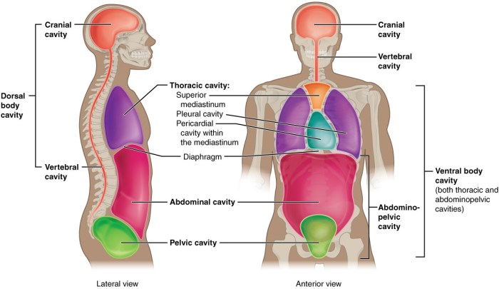
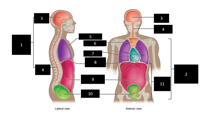
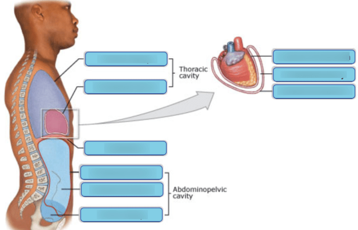
Art-labeling activity the anterior and posterior body cavities – Embark on an enlightening journey with the art-labeling activity, a captivating educational tool that illuminates the complexities of the anterior and posterior body cavities. This immersive experience empowers learners to visualize and comprehend the intricate structures within these cavities, fostering a deeper understanding of human anatomy.
Through the act of labeling anatomical structures, students engage in active learning, solidifying their grasp of the subject matter. This hands-on approach transforms passive knowledge into an interactive and memorable experience, enhancing retention and comprehension.
Art-Labeling Activity: Enhancing the Understanding of Anterior and Posterior Body Cavities: Art-labeling Activity The Anterior And Posterior Body Cavities

Art-labeling activities are an effective teaching tool that engages students in the process of learning by combining visual and kinesthetic elements. They provide a hands-on approach to understanding complex anatomical structures and their relationships.
For the study of the anterior and posterior body cavities, art-labeling activities are particularly beneficial as they allow students to visualize and interact with the structures within these cavities. By physically labeling and manipulating images, students can develop a deeper understanding of the location, shape, and relationships between these structures.
Anterior Body Cavity, Art-labeling activity the anterior and posterior body cavities
The anterior body cavity, also known as the ventral body cavity, is located in the front of the body. It contains the following structures:
- Heart
- Lungs
- Esophagus
- Trachea
- Thymus
- Pleural cavities (containing the lungs)
- Pericardial cavity (containing the heart)
- Mediastinum (separating the pleural cavities)
Art-labeling activities can enhance the visualization of these structures by providing students with a tangible representation of their location and relationships. By labeling and manipulating images of the anterior body cavity, students can gain a better understanding of the spatial arrangement of these structures and their relative sizes.
Posterior Body Cavity
The posterior body cavity, also known as the dorsal body cavity, is located in the back of the body. It contains the following structures:
- Stomach
- Small intestine
- Large intestine
- Liver
- Pancreas
- Gallbladder
- Kidneys
- Adrenal glands
- Urinary bladder
- Reproductive organs
- Peritoneal cavity (containing the digestive organs)
- Retroperitoneal cavity (containing the kidneys and adrenal glands)
Art-labeling activities can improve the understanding of the relationships between these structures by allowing students to physically connect and manipulate them. By labeling and manipulating images of the posterior body cavity, students can gain a better understanding of the relationships between the digestive, urinary, and reproductive systems.
Comparison of Anterior and Posterior Body Cavities
The following table compares the structures found in the anterior and posterior body cavities:
| Anterior Body Cavity | Posterior Body Cavity |
|---|---|
| Heart | Stomach |
| Lungs | Small intestine |
| Esophagus | Large intestine |
| Trachea | Liver |
| Thymus | Pancreas |
| Pleural cavities | Gallbladder |
| Pericardial cavity | Kidneys |
| Mediastinum | Adrenal glands |
| Urinary bladder | |
| Reproductive organs |
As can be seen from the table, the anterior and posterior body cavities contain different sets of organs and structures. The anterior body cavity is primarily associated with the respiratory and circulatory systems, while the posterior body cavity is associated with the digestive, urinary, and reproductive systems.
Educational Applications
Art-labeling activities can be incorporated into lesson plans in a variety of ways. They can be used as a pre-test to assess students’ prior knowledge, as a hands-on activity to reinforce concepts, or as a post-test to evaluate students’ understanding of the material.
For example, an art-labeling activity could be used to teach students about the structures of the anterior body cavity. Students could be given an image of the anterior body cavity and asked to label the different structures. Once students have labeled the structures, they could be asked to describe the relationships between them and explain how they work together.
Art-labeling activities can also be used to teach students about the relationships between the anterior and posterior body cavities. Students could be given an image of the entire body cavity and asked to identify the different structures in each cavity.
Once students have identified the structures, they could be asked to describe the relationships between them and explain how they work together.
Conclusion
Art-labeling activities are a valuable teaching tool for understanding the anterior and posterior body cavities. They provide a hands-on approach to learning that allows students to visualize and interact with the structures within these cavities. By labeling and manipulating images, students can develop a deeper understanding of the location, shape, and relationships between these structures.
Art-labeling activities can be incorporated into lesson plans in a variety of ways and can be used to teach students about the structures of the anterior and posterior body cavities, as well as the relationships between them.
User Queries
What is the purpose of an art-labeling activity?
Art-labeling activities are designed to enhance visualization and understanding of anatomical structures by actively engaging learners in the labeling process.
How does art-labeling improve comprehension?
The act of labeling requires students to identify and locate specific structures, reinforcing their understanding of their location and relationships within the body.
Can art-labeling activities be used for both anterior and posterior body cavities?
Yes, art-labeling activities can be effectively employed to study both the anterior and posterior body cavities, providing a comprehensive understanding of human anatomy.

