Exercise 13 review sheet neuron anatomy and physiology – Exercise 13 Review Sheet: Neuron Anatomy and Physiology delves into the intricate world of neurons, the fundamental units of our nervous system. This comprehensive resource provides a thorough understanding of neuronal structure and function, paving the way for a deeper exploration of neurophysiology and its clinical applications.
Delving into the intricacies of neuron anatomy, we examine the cell body, dendrites, and axon, deciphering their specialized roles in receiving, processing, and transmitting electrical signals. The detailed diagram provided serves as a visual guide, aiding in the comprehension of these vital components.
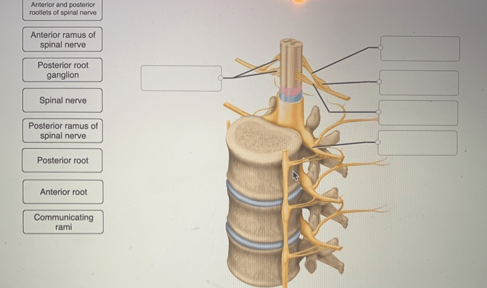
Neuron Anatomy

Neurons are the fundamental units of the nervous system. They receive, process, and transmit information throughout the body. Each neuron consists of a cell body, dendrites, and an axon.
The cell body, or soma, contains the nucleus and other organelles responsible for the neuron’s metabolism and protein synthesis. Dendrites are short, branched extensions that receive signals from other neurons. The axon is a long, slender projection that transmits signals away from the cell body to other neurons or muscles.
Neurons are highly specialized cells with unique structural and functional properties that enable them to perform their vital role in communication and information processing within the nervous system.
Neuron Physiology
The resting membrane potential of a neuron is a stable electrical potential difference across the neuron’s plasma membrane. It is maintained by the differential distribution of ions across the membrane and the activity of ion channels.
When a neuron receives a sufficiently strong stimulus, it undergoes an action potential. An action potential is a brief, all-or-nothing electrical pulse that travels along the axon. It is generated by the opening and closing of voltage-gated ion channels in the axon membrane.
Ion channels are proteins embedded in the neuron’s plasma membrane that allow specific ions to pass through. The opening and closing of ion channels are controlled by the neuron’s electrical potential and by chemical signals from other neurons.
Synaptic Transmission
Synapses are specialized junctions between neurons where signals are transmitted from one neuron to another. They consist of a presynaptic terminal, a synaptic cleft, and a postsynaptic terminal.
When an action potential reaches the presynaptic terminal, it triggers the release of neurotransmitters into the synaptic cleft. Neurotransmitters are chemical messengers that bind to receptors on the postsynaptic terminal, causing a change in the electrical potential of the postsynaptic neuron.
Synaptic transmission can be either excitatory or inhibitory. Excitatory synapses cause the postsynaptic neuron to become more likely to fire an action potential, while inhibitory synapses make it less likely.
Neuroanatomy and Neurophysiology Techniques
A variety of techniques are used to study neuron anatomy and physiology. These techniques include electrophysiology, patch clamping, and immunohistochemistry.
Electrophysiology involves recording the electrical activity of neurons using electrodes. Patch clamping is a technique that allows researchers to record the activity of single ion channels.
Immunohistochemistry is a technique that uses antibodies to visualize specific proteins in neurons.
Clinical Applications of Neuroanatomy and Neurophysiology, Exercise 13 review sheet neuron anatomy and physiology
Neuroanatomy and neurophysiology have a wide range of clinical applications. They are used in the diagnosis and treatment of neurological disorders, such as epilepsy, Parkinson’s disease, and Alzheimer’s disease.
Neuroanatomy and neurophysiology can also be used to develop new therapies for neurological disorders. For example, deep brain stimulation is a surgical procedure that uses electrodes to stimulate specific brain regions to treat movement disorders such as Parkinson’s disease.
Question Bank: Exercise 13 Review Sheet Neuron Anatomy And Physiology
What is the primary function of dendrites?
Dendrites receive electrical signals from other neurons and transmit them to the cell body.
How is an action potential generated?
An action potential is generated when the influx of sodium ions into the neuron exceeds the efflux of potassium ions, causing a rapid depolarization of the membrane.
What is the role of neurotransmitters in synaptic transmission?
Neurotransmitters are chemical messengers that are released from the presynaptic neuron and bind to receptors on the postsynaptic neuron, influencing its electrical activity.